石上先生がDigestive Disease Week 2018 (June 2-5, 2018@Washington D.C.) に参加しました
6/2-5の日程で米国Washington D.C.で開催されていた、Digestive Disease Week 2018に参加し、poster presentationを行いました。臨床検体でご協力頂いた市立室蘭総合病院・金戸宏行先生、本学病理診断部の長谷川匡教授、消化器外科・竹政伊知朗教授、木村康利先生に御礼申し上げます。また、何よりご指導いただいた仲瀬裕志教授、能正勝彦先生のおかげでこのような機会が得られたと思っています。
お世話になったすべての先生に、この場をお借りして深く御礼申し上げます。
昨年は緊張して周りが見えませんでしたが、今年は自分のポスターの前を素通りする人ばかりで、非常に悔しい思いをしました。次はもっともっと面白いデータを持って、先輩や後輩と一緒に参加したいなぁと思います。
弾丸ツアーだったので観光はほとんどできず、行きかえりのタクシーの運転手との会話(片道約40分)が一番現地の人とたくさんしゃべった思い出です。
英語もやはり日々の鍛錬が重要だな、と痛感しています…。
MicroRNA-31 reflects IL-6 expression in cholangiocarcinoma tissue and is related to patient survival
Background & Aims: We previously reported the impact of MicroRNA-31 (miR-31) on patient survival in gastrointestinal cancers (e.g., colorectal cancer and pancreatic cancer). With regard to cholangiocarcinoma, miR-31 has been reported to be upregulated in tumor tissues, and it not only promotes cellular proliferation but also inhibits apoptosis. Nevertheless, no study has reported the association between miR-31 expression and patient survival. We therefore evaluated miR-31 expression in cholangiocarcinoma tissues and its relationship with prognosis. Furthermore, we examined the impact of several cytokines on miR-31 expression in cholangiocarcinoma cell lines to investigate whether this could reflect cytokine expression in tumor tissues.
Methods: To examine the expression of miR-31 in cancer tissues, we performed quantitative reverse transcription-polymerase chain reaction in 81 cholangiocarcinoma patients (16 with intrahepatic cholangiocarcinoma, 37 with extrahepatic cholangiocarcinoma, and 28 with gallbladder cancer). To investigate the influence of cytokines on miR-31 expression, cholangiocarcinoma cell lines were stimulated with cytokines. For functional analysis, we conducted proliferation assays and Western blotting.
Results: The expression level of miR-31 was significantly higher in cholangiocarcinoma cells than in normal bile duct epithelium cells (P = 0.038). A significant association was not detected between miR-31 expression and clinical or pathological characteristics, such as gender, age, tumor location, N factor, M factor, and disease stage, except for tumor size (P = 0.012). In the Kaplan–Meier analysis, high miR-31 expression was significantly associated with shorter survival (log-rank test, P = 0.0082). In the multivariate Cox regression analysis, high miR-31 expression was significantly associated with prognosis (P = 0.043), independent of clinical and pathological features. Functional analysis of cholangiocarcinoma cell lines revealed that IL-6 significantly promoted miR-31 expression and cell proliferation in a dose-dependent manner. The promoter of miR-31 contains a potential binding site for phosphorylated STAT-3, and inhibition of STAT-3 signaling significantly suppressed miR-31 expression and cell proliferation.
Conclusion: High miR-31 expression was observed in cholangiocarcinoma cells, and this high expression was significantly associated with worse prognosis in the patients. Our data also revealed that the IL-6–STAT-3 signaling pathway regulates the proliferation of cholangiocarcinoma cells and the expression of miR-31. These data suggest that miR-31 may be a promising biomarker that reflects IL-6 expression in cholangiocarcinoma tissues and predicts worse prognosis.
 トップページ
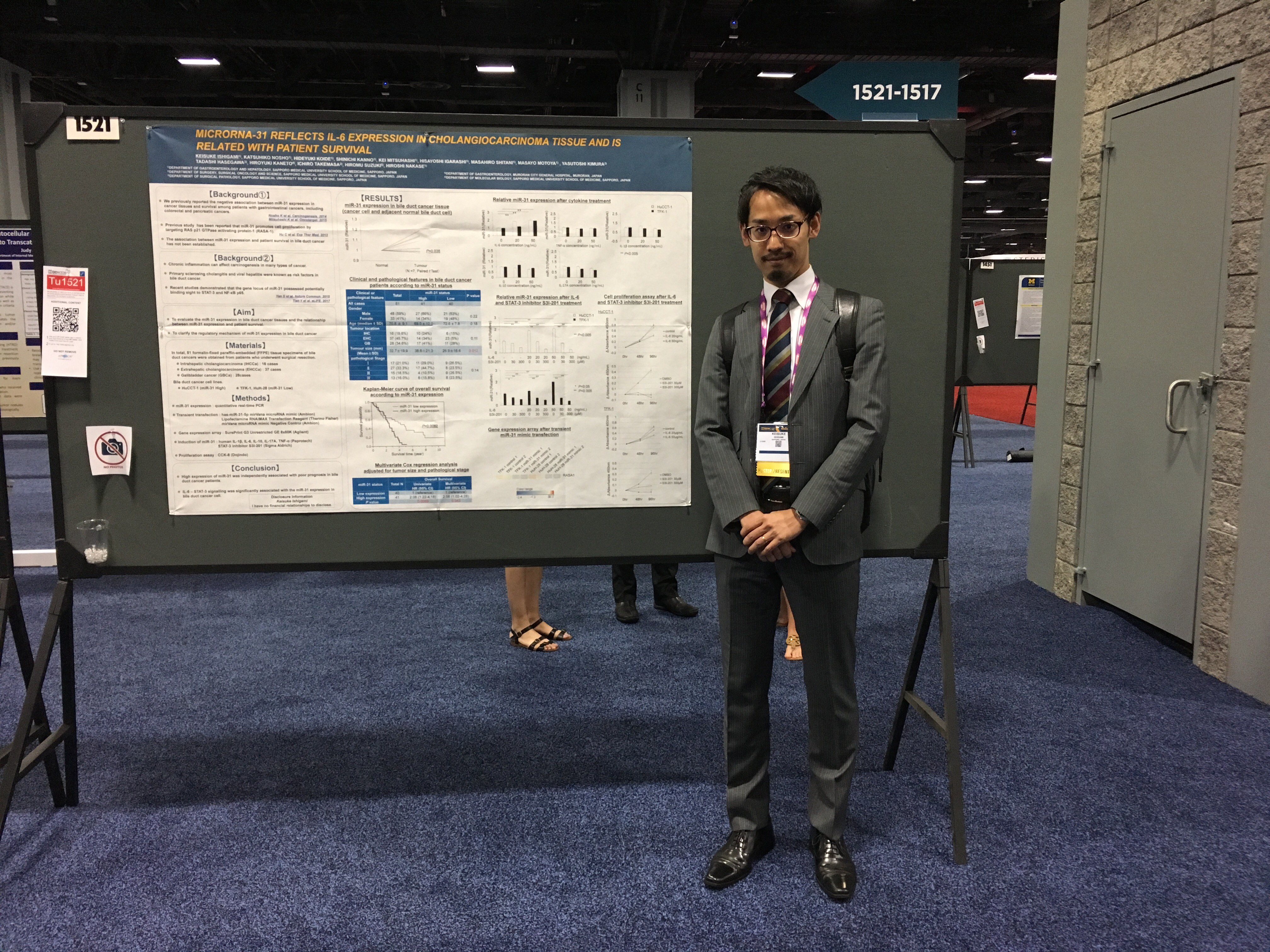